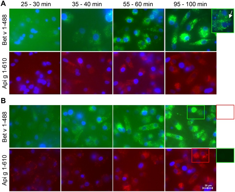
Fig 1. Kinetics of allergen uptake into iMoDCs of BP allergic and normal donors.
Internalization of labeled Bet v 1 (Bet v 1–488) and labeled Api g 1 (Api g 1–610) by iMoDCs of allergic (A) and normal donors (B) was followed by live-cell fluorescence imaging. Results are representative of independent experiments of three different donors for each group (shown for donors AD1 and ND3). Exocytosis of fluorescent markers (panel A) and the absence of spillover of fluorescence (panel B) are depicted by framed display details.

