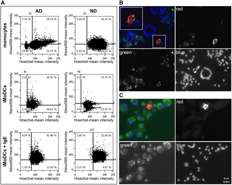
Fig 7. Binding of IgE and Bet v 1 to monocytes and iMoDCs of BP allergic (AD) and normal donors (ND).
Cells were grown in chamber slides and stained with Hoechst33342 to label the nuclei. Surface-bound IgE was detected with primary antibodies against IgE and Alexa Fluor 568-labeled secondary antibodies. (A) Quantification of surface-bound IgE and IgE receptors using a TissueFAXS microscopy system. FcεRI surface expression was determined indirectly by loading iMoDCs additionally with 1μg/ml human myeloma IgE (iMoDCs + IgE). Results represent data typical for two independent experiments for each donor group (shown for donors AD2 and ND2). Percentage of IgE-positive cells is indicated in the upper right corner of the scattergram. (B) Fluorescence microscopy of iMODCs of BP allergic and (C) normal donors. Cells were incubated with Alexa 488-labeled Bet v 1 (green) and Alexa Fluor 568-labeled anti-IgE antibodies (red). The magnified insert in (B) shows an individual cell with non-overlapping staining of Bet v 1 and IgE.

