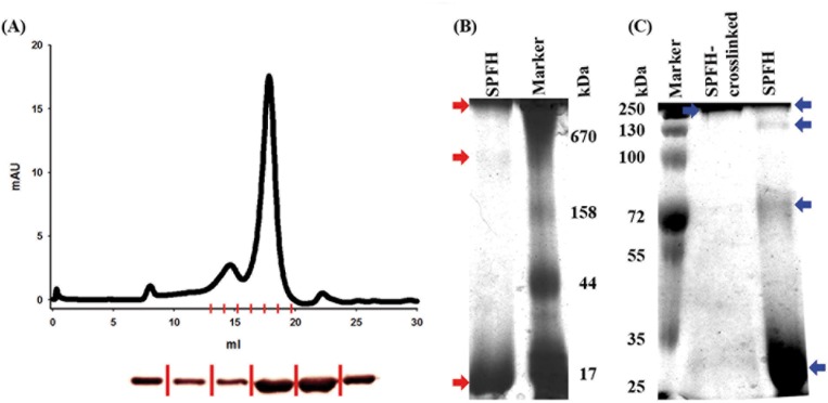
Figure 4. Oligomerization of the PHB domain.
(A) Size exclusion chromatography was performed with the PHB domain on a Superose6 column (GE Healthcare) and two elution peaks were detected at 14.6 and 17.5 ml, corresponding to a size of 313 kDa and 42.8 kDa respectively. (B) Blue native PAGE was performed with the PHB domain, three bands marked with red arrows could be detected corresponding to a monomer, a 14mer (313 kDa) and an oligomer larger than 670 kDa. (C) A colloidal Coomassie blue stained SDS—PAGE gel is shown. The PHB domain was further probed by cross-linking and applying the sample to SDS-PAGE. For cross-linked PHB only a band larger than 250 kDa is detected, for none cross-linked PHB domain bands corresponding to a monomer, a tetramer, an octamer and an oligomer larger than 250 kDa are be detected (indicated by blue arrows). The SEC column was calibrated using standard proteins (see S2 Fig.).

