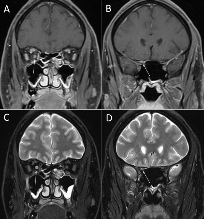
Figure 1. Gd enhancement and T2 lesion of the right optic nerve.
Optic nerve MR imaging of a representative patient with ON. (A) Coronar fat-saturated T1-weighted MRI sequences of the intraorbital and (B) canalicular part of the optic nerve after application of 0.1mmol/kg gadolinium. The arrows highlight the Gd enhanced right optic nerve. (C) Signalalteration of the intraorbital and (D) canalicular part of the optic nerve in coronar fat-saturated T2 turbo spin-echo MRI sequences. The arrows highlight the hypertense T2 lesion of the right optic nerve.

