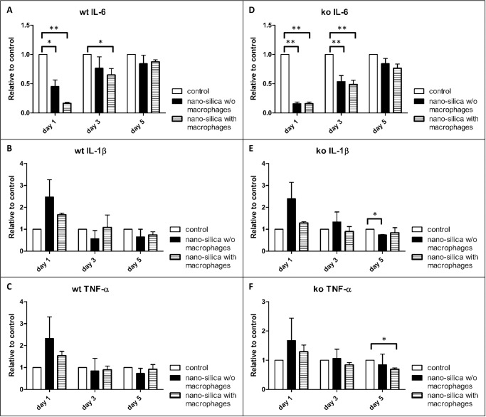
Figure 9. Measurement of IL-6 (A, D), TNF-α (B, E) and IL-1β (C, F) in the supernatant of the PCLS via ELISA.
Compared are wild type (wt) and P2rx7−/− (ko) mice that were cultivated with and without (w/o) AMJ2-C11 macrophages with a nano-silica concentration of 1600 µg/ml. Data is presented in relation to the controls. The incubation time was 72 hours. n = 6–9.

