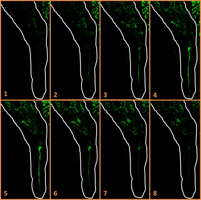
Figure 3. Localization of Y. enterocolitica YE03 in Steinernema sp. MW8B EPNs emerged from an infected larva.

Confocal microscope slides in Z-axis (numbered from 1 to 8) of a Steinernema sp. MW8B EPN colonized by Y. enterocolitica YE03 emerged from the second infection cycle. GFP-labeled bacteria localize in the mouth and in the gut lumen. EPN borders are drawn in white (800× magnification).
