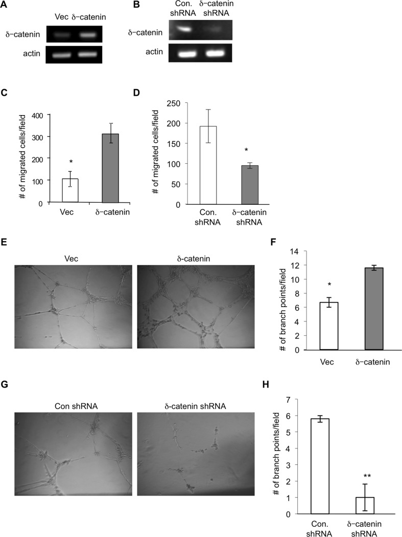
Figure 1. δ-catenin is present in lymphatic endothelial cells and regulates lymphangiogenesis in vitro.
HMVEC-LLy cells were transfected with either an empty vector (vec) or an expression vector for δ-catenin for 48 hours. Total RNA was isolated and δ-catenin mRNA levels were quantitated by semi quantitative RT-PCR (Panel A). HMVEC-LLy cells were either transfected with a mixture of δ-catenin specific shRNA constructs or control shRNA construct for 48 hours, respectively. Total RNA was isolated and δ-catenin mRNA levels were quantitated by semi quantitative RT-PCR (Panel B). Cell migration was measured in a Transwell chamber by seeding HMVEC-LLy cells transfected with empty vector or δ-catenin expression vectors. Migrated cells were counted from 10 randomly selected high power fields under microscopy 4 hours after incubation and graphed with mean and standard error (Panel C) *p<0.05. Cell migration was also measured by seeding δ-catenin knockdown HMVEC-LLy cells or control shRNA transfected cells for 4 hours, followed by the same procedure as described above (Panel D) *p<0.05. HMVEC-LLy cells transfected with either a vector control or δ-catenin expression vector were incubated on Matrigel for 24 hours. The images of vascular network formation were taken under microscopy (Panel E). The number of vascular branch points was counted in 10 randomly selected fields under microscopy (Panel F). Vascular network formation in Matrigel was evaluated using δ-catenin knockdown HMVEC-LLy cells and control cells (Panel G and H). Representative images were shown. *p<0.05; **p<0.01. Each experiment was done in triplicate and the experiment was repeated twice.

