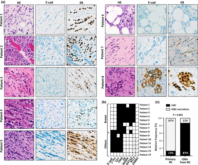
Figure 2.
(A) Representative photomicrographs of orbital metastases (OMs) from patients with a history of breast cancer. HE-stained sections are shown on the left side (400×), immunohistochemical stainings for E-cadherin (E-cad) and estrogen receptor (ER) are shown on the right side. (B) Overview showing the immunophenotypical characteristics of the 14 OM. Immunohistochemical markers are aligned along the bottom, primary tumor sites along the left side. Filled squares indicate positive staining. Empty squares indicate negative staining. Diagonal lines indicate missing data. (C) Proportion of infiltrating lobular breast cancer (ILBC) in a control group of randomly selected primary breast cancers (BC, n = 17/134) diagnosed at our institution, compared with the proportion of ILBC in OMs from patients with a history of breast cancer (n = 7/8). Statistical significance was assessed with the chi-square test using the actual numbers of observed cases.

