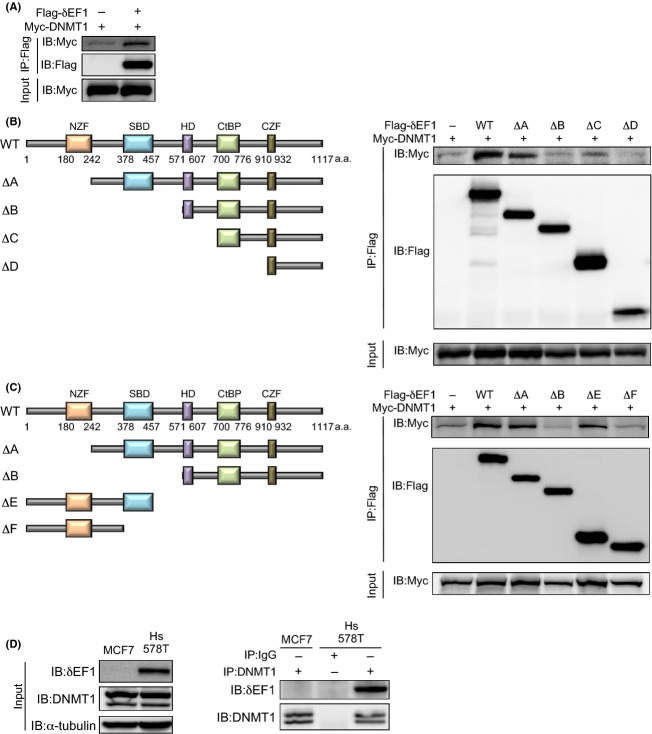
Figure 3.
Interaction of δEF1 with DNMT1. (A–C) HEK293 cells were transiently transfected with the indicated expression plasmids. Twenty-four hours after transfection, cells were harvested, lysed, and subjected to immunoprecipitation (IP) with anti-FLAG antibody, followed by immunoblotting (IB) with anti-Myc antibody. Schematic illustrations depict wild-type (WT), N-terminally truncated mutants (ΔA–ΔD), and C-terminally truncated mutants (ΔE–ΔF) of δEF1 (left panels in B and C). (D) MCF7 and Hs578T cells were harvested and subjected to immunoprecipitation (IP) with anti-DNMT1 antibody or IgG, followed by immunoblotting (IB) with anti-DNMT1 or anti-δEF1 antibodies. α-tubulin levels were monitored as a loading control. NZF, N-terminal zinc finger; SBD, Smad-binding domain; HD, homeodomain; CtBP, CtBP-binding domain; CZF, C-terminal zinc finger.

