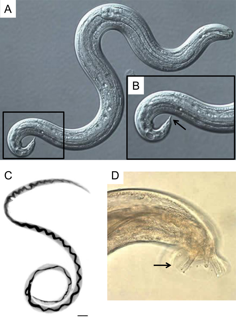
Figure 1.
Angiostrongylus cantonensis developmental stages. (A) Differential interference contrast microscopy image of third-stage (L3) infective larvae recovered from a slug. L3 larvae are about 0.45 by 0.02 mm and present cuticle with faint transverse striations. (B) Higher magnification of demarcated region in A showing terminal projection on the tip of the tail (arrow) which is characteristic of A. cantonensis. Adult female (C) and tail of adult male (D) worms recovered from rat lungs. Note the characteristic barber-pole appearance of female worms and rays (arrow) of males. Scale bar = 1 mm in C. Sources: (A), (B) and (D) = http://www.cdc.gov/dpdx/angiostrongyliasis/gallery.html; (C) = http://wwwnc.cdc.gov/eid/article/8/3/01-0316-f1.htm.

