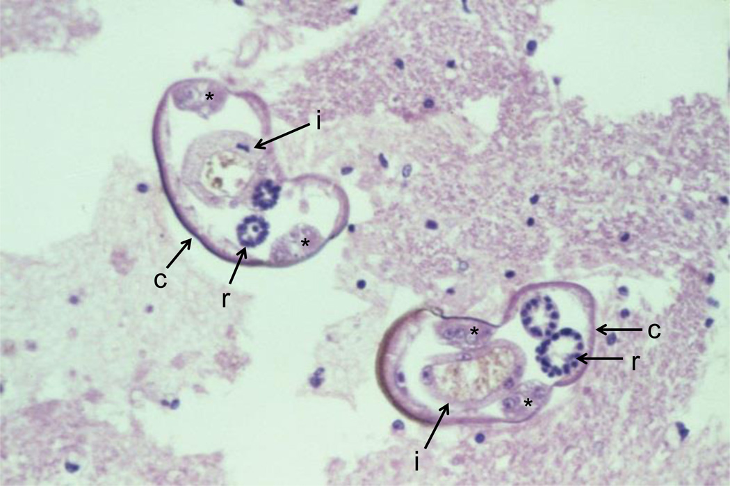
Figure 2.
Cross-section of two Angiostrongylus cantonensis larvae in the spinal cord. Note the smooth thin cuticle (c), lateral cords (*), intestines (i) with few multinucleated cells and reproductive tubes (r). Source: http://www.astmh.org/source/ZaimanSlides/index.cfm?photo=BEA3A648-E0C7-F0E4-AAEC8C1C0EB551C5.

