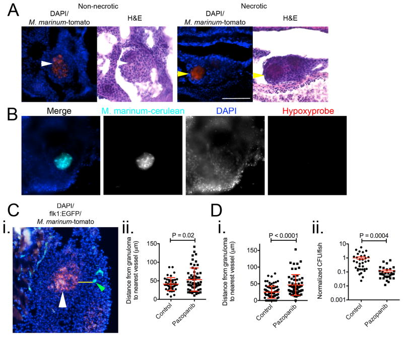
Extended Data 6.
(A) Images of non-necrotic (left) and necrotic (right) M. marinum-tomato granulomas stained with DAPI (top) and haematoxylin & eosin (bottom). White arrows indicate non-necrotic granuloma, yellow arrows indicate necrotic granulomas. Images are representative of granulomas found in 8 individual animals.
(B) Representative image of a necrotic granuloma from a negative control, not injected with pimonidazole, 2 wpi adult Tg(flk1:EGFP) zebrafish infected with M. marinum-cerulean (cyan), and stained for hypoxyprobe (red) and with DAPI (blue). Images are representative of granulomas found in 2 individual animals.
(C) i. Representative image of M. marinum-tomato granuloma in Tg(flk1:EGFP) zebrafish stained with DAPI. White arrow indicates granuloma, yellow line indicates path measured for distance between granuloma and nearest vasculature (indicated by green arrow). Image is representative of data presented in Extended Data 6Cii, Extended Data 6Di and Extended Data 7A. ii. Distance between granulomas and nearest vasculature measured in 2 wpi adult Tg(flk1:EGFP) zebrafish. Total number of zebrafish analysed = 4 (control), 4 (pazopanib).
(D) i. Distance between granulomas and nearest vasculature measured in 2 wpi adult Tg(flk1:EGFP) zebrafish treated with pazopanib for 1 week. Total number of zebrafish analysed = 2 (control), 2 (pazopanib). ii. Bacterial burden in 2 wpi adult zebrafish treated with pazopanib for 1 week. T-test, data are pooled from 3 biological replicates.

