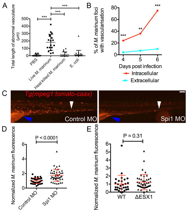
Extended Data 2.
(A) Length of abnormal vasculature in Tg(flk1:EGFP) larvae injected with PBS, live M. marinum, heat killed M. marinum and E. coli. One-way ANOVA with Tukey’s post-test, data are representative of two biological replicates.
(B) Recruitment of vasculature by intracellular and extracellular foci of M. marinum. Total number of foci analysed: 4 dpi 221 intracellular 105 extracellular, 5 dpi 71 intracellular 26 extracellular and 6 dpi 131 intracellular 50 extracellular. Fisher’s exact test.
(C) Comparative images of 5 dpf control and Spi1 morphant Tg(mpeg1:tomato-caax)xt3 larvae. White arrowhead indicates comparative locations within the caudal haematopoietic tissue. Blue arrowhead indicates intestinal and yolk sac autofluorescence. Scale bar indicates 100 μm. Images are representative of transgene expression in 20 animals per treatment group.
(D and E) Bacterial burden in: (D) 5 dpi control and Spi1 morphant larvae, and (E) 4 dpi larvae infected with WT or ΔESX1 M. marinum-tomato. T-test with Welch’s correction, all data are pooled from two biological replicates.
Error bars represent mean ± standard deviation.

