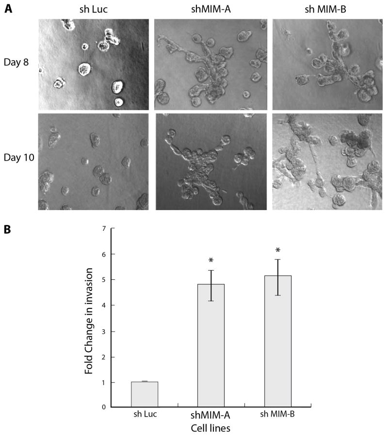
Figure 2. Suppression of MIM promoted mammary epithelial cell invasion.
(A) Induction of invasive structures in MCF10A cells in which MIM was suppressed by two distinct shRNAs. The micrographs were taken at Day 8 and Day 10 of 3D culture in Matrigel-collagen. Scale bar represents 100 μm. (B) MCF10A cells infected with control or two distinct MIM-directed shRNAs were seeded in transwell migration chambers in which the membrane was coated with Matrigel, incubated for 48 h, and cells that had migrated through the Matrigel coating were counted. Invasion by the control cells was normalized to 1. Error bars represent S.E.M. (n=3). * denotes p value<0.05

