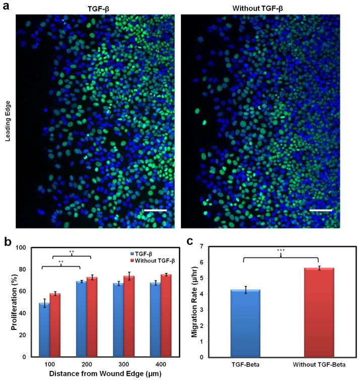
Fig. 5.
Proliferation of migrating MCF-7 cells was measured in a wound healing assay. Proliferation was compared with and without the addition of TGF-β. Over the course of twenty-four hours proliferation inhibition occurred regardless of the vicinity of the cells to the leading edge of the wound. (a) Fluorescence images demonstrating the proliferation measurements. MCF-7 cells were dyed with Hoechst 33342 (blue) and Alexa Fluor 488 (green) via the Click-iT EdU Imaging Kit (Invitrogen). Alexa Fluor 488 presents cells that synthesized DNA over the course of twenty-four hours, while Hoechst 33342 highlights the total number of cells overall. The scale bars are 100 μm. (b) Graph demonstrating the spatial proliferation difference between cells with and without TGF-β addition (**, P<0.01). Cells near the wound edge proliferated less than cells in the monolayer, regardless of TGF-β addition. (c) Collective migration of MCF-7 cells in a wound healing assay with and without TGF-β addition. MCF-7 cells grown to confluence were scratched with a pipette tip; this allowed the cells to migrate into the wound area. Migration rate was determined after twenty-four hours of migration by measuring the area covered by the cells over that time and dividing by the width of the monolayer (***, P<0.001).

