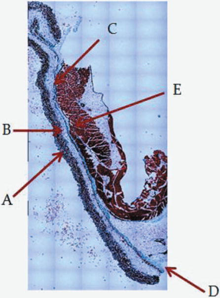
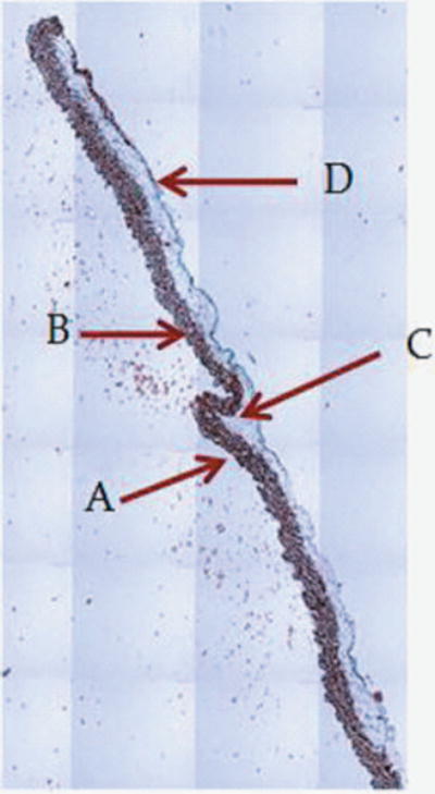
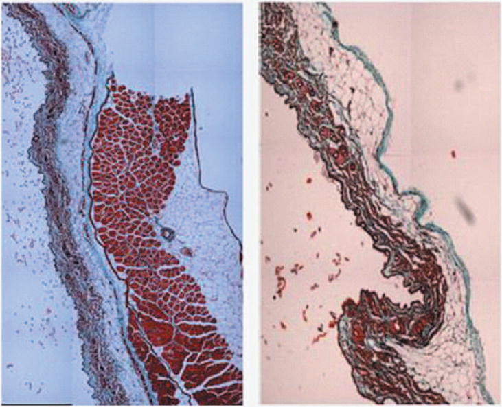
Figure 1.


1A: 5X magnification of skin and abdomen samples. On the left Trichrom staining of the abdominal intact wall-skin samples obtained from HA mice. On the right is the skin sample obtained after a simulated sc injection using the “pinch” method. This figures show that the “pinch” method is separating the skin at the membranous layer, which results in a much deeper deposit of the sc dose than expected in humans. Arrow are pointing at: A: Dermis. B: Epidermis. C: Superficial adipose tissue D: Deep adipose tissue E: Membranous layer. 1B: 40X magnification of skin and abdomen samples. On the left Trichrom staining of the abdominal wall-skin samples obtained from HA mice we can see the panniculus carnosus (red intermittent muscular layer). On the right is the skin sample obtained from the “pinch” method. Here we can see the membranous layer (blue) appearing thinner when compared to the image to the right.
