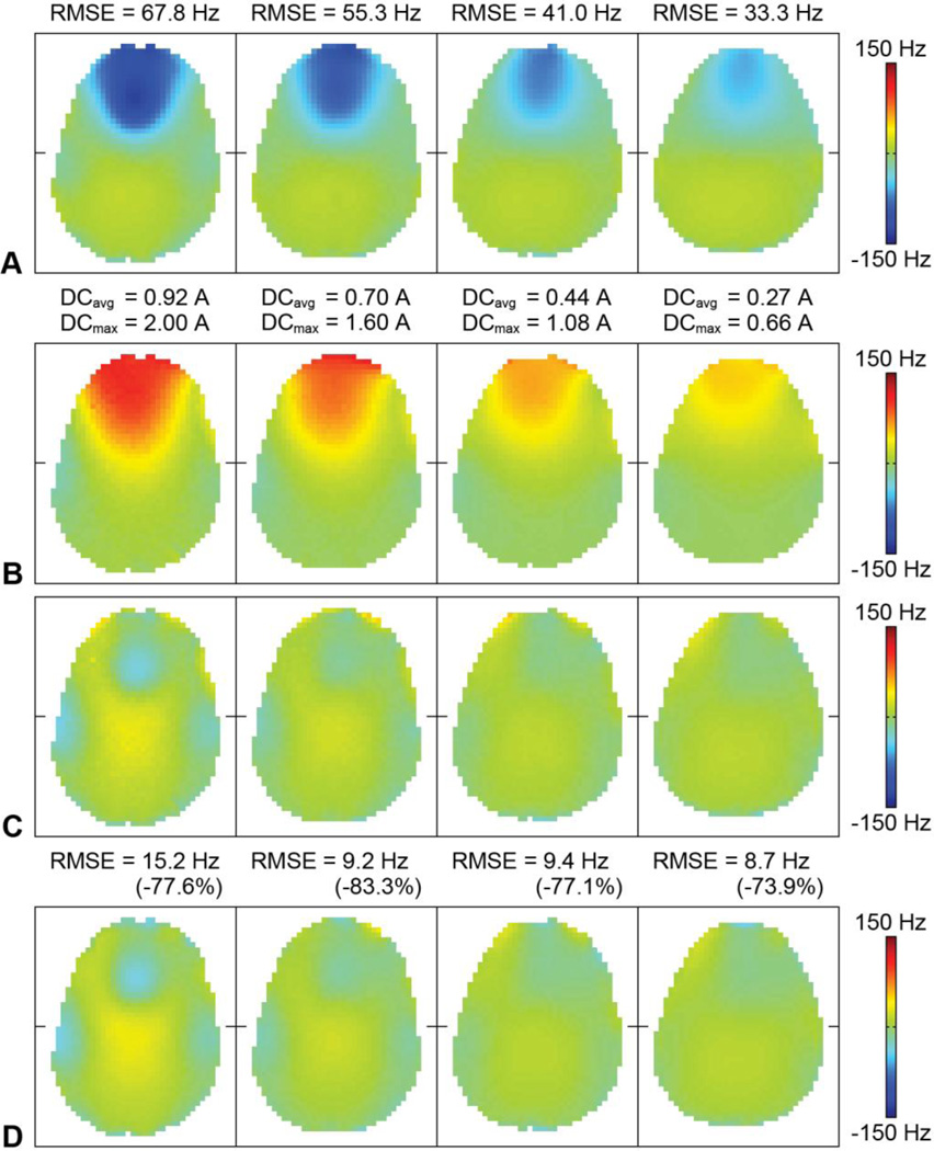
Fig. 4.
(A) B0 maps acquired in vivo without DC currents in four representative slices. The B0 RMSE in the anterior half of the brain is shown at the top. (B) B0 field generated by the RF/shim coils with optimal DC currents (i.e., sum of the basis B0 maps weighted by the optimal DC currents). The average and maximum DC current amplitudes applied in the RF/shim coils are shown at the top. (C) B0 maps with optimal DC currents predicted from the shim optimization (i.e., sum of (A) and (B)). (D) B0 maps acquired in vivo with optimal DC currents. The B0 RMSE in the anterior half of the brain and percent reduction with respect to (A) are shown at the top.

