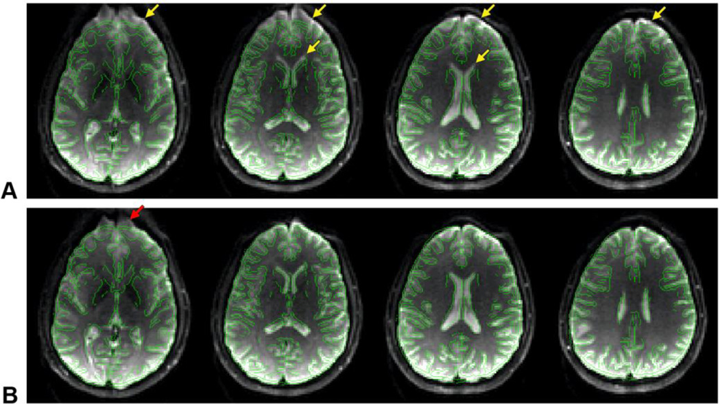
Fig. 5.
EPI images acquired in the same slices as in Fig. 4 without DC currents (A) and with optimal DC currents (B), with overlaid contour lines derived from the undistorted fast spin-echo images. The arrows point to susceptibility-induced distortions in the frontal brain region.

