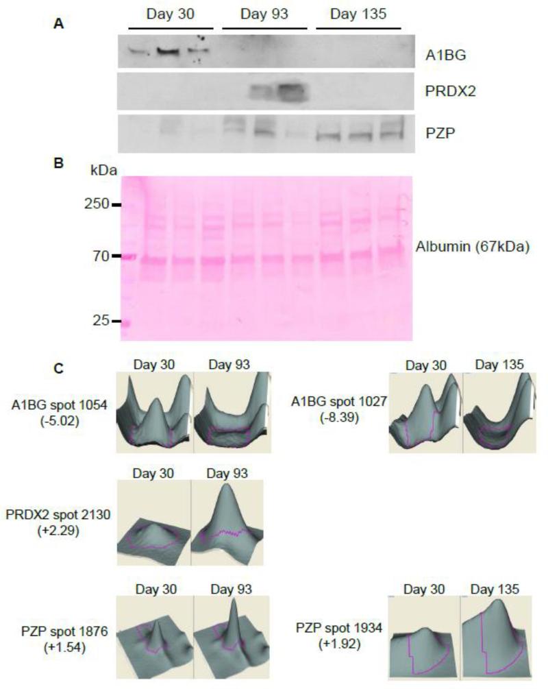
Figure 4. Western blot analysis of putative IBD biomarkers A1BG, PRDX2, and PZP, and corresponding 3D view.
A, Accumulation of A1BG, PRDX2, and PZP in serum samples from mice with mild/moderate or severe colitis compared to that in serum from non-colitic mice was validated by Western blotting. B, Ponceau Red-stained Western blot membrane, used as a loading control. Albumin is visible at 67 kDa. C, 3D rendering of A1BG, PRDX2, and PZP protein spots enlarged to show the differences in their expression between non-colitic mice (30 days old) and mice with moderate (93 days old) and severe (135 days old) colitis. The 3D views of 1 out of 3 independent experiments are shown.

