
Figure 3.
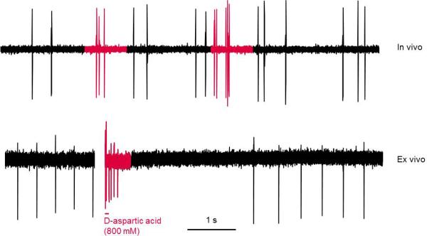
Dopamine neurons burst differently between in vivo and ex vivo preparations. Top panel: representative trace showing spontaneous bursting of a dopamine neuron in vivo in a chloral hydrate anesthetized rat. Bottom panel: representative trace showing bursting of a dopamine neuron recorded in cell-attached configuration in the ex vivo tissue slice preparation. Bursting was induced by iontophoretic application of D-aspartic acid in the presence of glycine in the perfusion bath. In vivo, firing activity consists of irregular single spikes interspersed with short (2-5) spike bursts. In contrast, in ex vivo slices dopamine neurons fire in an extremely regular pacemaker-like fashion with induced bursts consisting of many spikes and being followed by a long post-burst pause. In vivo trace collected in our lab; ex vivo trace provided by Dr. Michael Beckstead.