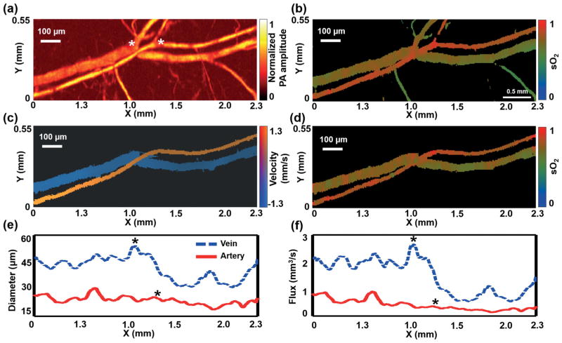
Figure 3.
For in vivo measurement, a 2.3 × 0.55 mm2 region in the ear of a living mouse was imaged by OR-PAM with raster and 3-DAT scanning. Global raster scanning acquired (a) a structural image and (b) a sO2 map of the main vascular trunk. The artery-vein pair was selected as the region of interest to guide 3-DAT scanning, yielding (c) the blood flow speed, (d) the sO2 map, (e) the diameter, and (f) the flow flux. The asterisks indicate the locations of bifurcations.

