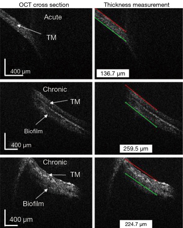
Figure 9.

In vivo acute and chronic OM imaging. Shown are in vivo OCT cross sections with the corresponding thickness measurements for one acute and two chronic OM cases. Inflammation in the acute case and the possible presence of biofilm in the chronic cases resulted in varying thickness measurements. OM, otitis media; OCT, optical coherence tomography; TM, tympanic membrane
