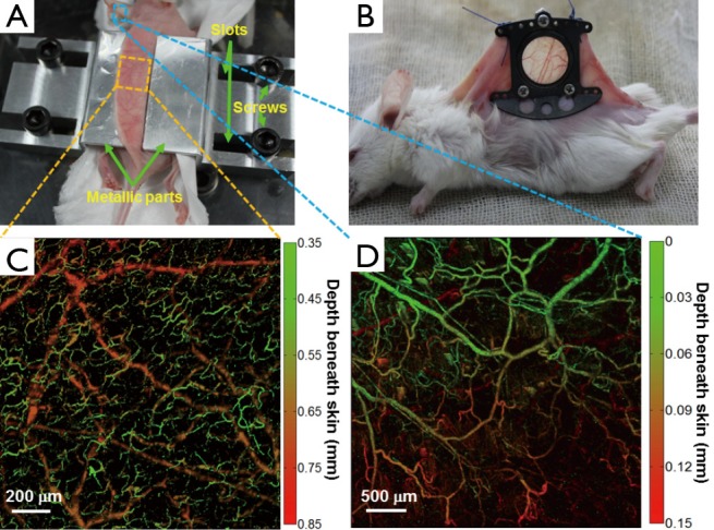
Figure 2.

(A) N-DSF mouse fixture designed for our OR-PAM system. The dorsal skin only needs to be slightly clamped temporarily using this fixture; (B) Traditional invasive skin-fold window chamber used for intravital optical microscopy, permanently mounted on the mouse back by a surgical operation; (C) a representative depth-encoded MAP image of a normal mouse’s dorsal region acquired by the combined use of OR-PAM and N-DSF fixture; (D) a representative depth-encoded MAP image of a normal mouse ear acquired by OR-PAM. The color scale represents depth below the skin surface. N-DSF, noninvasive dorsal skin-fold; OR-PAM, optical-resolution photoacoustic microscopy; MAP, maximum amplitude projection.
