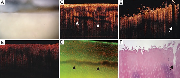
Figure 4.

Arthroscopic OCT imaging of human knee cartilage samples that display chondromalacia, uniformly graded 0/1 by conventional surface imaging. OCT is able to provide additional discrimination between these samples, with image B revealing an intact surface whilst C shows subsurface clefts and E shows fissures and fibrillations. Reproduced from Chu et al. (15) with permission. OCT, optical coherence tomography.
