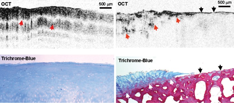
Figure 5.

In vivo polarization-sensitive OCT imaging of human knee cartilage, taken during knee replacement surgery. Upper row shows PS-OCT images, lower row shows contemporary histology, with Masson trichrome staining for collagen. Normal knee cartilage displays a uniform pattern of banding, suggesting a thick layer of organized birefringent collagen fibers. Exposed bone lacks obvious banding, as does degraded cartilage in the vicinity of the lesion. Reproduced from Li et al. (24) with permission. OCT, optical coherence tomography; PS-OCT, polarization-sensitive optical coherence tomography.
