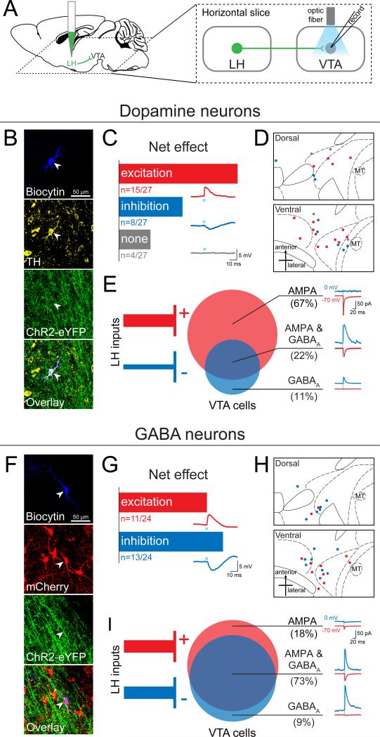
Figure 6. The LH sends a mixture of excitatory and inhibitory projections to both dopamine (DA) and GABA neurons in the VTA.
(A) AAV5-CaMKIIα-ChR2-eYFP was injected into the LH and at least 6 weeks later 300 μm thick horizontal brain slices were prepared containing the VTA. Whole-cell patch-clamp recordings were made in VTA neurons, and ChR2-expressing LH terminals were activated by illumination with 473 nm light via an optic fiber resting on the brain slice. (B) Neurons were filled with biocytin during recording, and DA neurons were identified by immunohistochemistry for tyrosine hydroxylase (TH) (n=27). (C) The net effect of optical stimulation of LH terminals was assessed in current-clamp mode, which revealed that 55% of DA neurons (n=15/27) showed a net excitatory response, while 30% (n=8/27) responded with net inhibition, and 15% (n=4/27) showed no response. An example of an excitatory postsynaptic potential (EPSP, red trace), an inhibitory postsynaptic potential (IPSP, blue trace), and a non-responsive cell (grey trace) are shown below each bar. (D) The distribution of all recorded TH+ neurons plotted on horizontal midbrain slices with colors indicating the response to LH terminal photostimulation. (E) VTA DA neurons received only AMPAR-mediated input (67%, n=6/9), only GABAAR-mediated input (11%, n=1/9) or both of these currents (22%, n=2/9). (F) VTA GABA neurons were identified by the presence of mCherry (n=24), achieved by injection of Cre-dependent AAV5-EF1α-DIO-mCherry into the VTA of VGAT::Cre mice. (G) Optical stimulation of LH terminals in current-clamp mode showed that GABA neurons respond with either net excitation (46%, n=11/24) or net inhibition (54%, n=13/24) to LH input. (H) The distribution of each recorded GABA neuron plotted on horizontal midbrain slices with colors indicating the response to LH terminal stimulation. (I) The monosynaptic input to VTA GABA cells recorded in the presence of TTX and 4AP revealed that GABA neurons receive a mixture of AMPAR-mediated and GABAAR-mediated input from the LH (AMPA only: 18%, n=2/11; AMPA & GABAA: 73%, n=8/11; GABAA: 9%, n=1/11). MT=medial terminal nucleus of the accessory optic tract. See also Figure S5 and S6.

