
Figure 3.
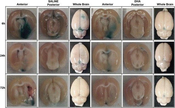
Representative images of EB extravasation from rats treated with DHA or saline. Treatments were given at 1 h after 2 h of MCAo and EB was administered at 1 h before rats were sacrificed (at 5 h, 23 h and 71 h). Extensive extravasation of EB is grossly visible in the saline treated rats. Contrarily, substantial inhibition of the dye extravasation was evident in DHA-treated brains.