
Figure 1.
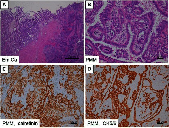
Hematoxylin and eosin (H&E) stains and immunohistochemical studies for two primary cancers. (A) H&E stain; endometrial cancer. (B) H&E stain; peritoneal malignant mesothelioma (PMM). High expression of (C) calretinin and (D) CK5/6 is shown in brown color in PMM. Scale bars represent 500 μm in (A), 50 μm in (B), and 100 μm in (C & D).