Figure 1.

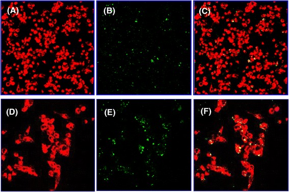
Uptake and internalization of VCGs by JAWS II dendritic cells (DCs). Internalization of fluorescence-labeled VCGs by JAWS II DCs and localization of VCGs within DCs were visualized by confocal scanning microscopy. DCs were incubated on Chamber slides for 45 min with 100 μg/ml of Alexa 594-labeled VCGs. Following extensive washing with PBS-BSA, cells were fixed in cold acetone and incubated with biotin-labeled anti-mouse CD11c antibodies and streptavidin-conjugated Alexa 488. Images were captured with a Leica scanning microscope with a 40x (A-C) or 63x oil (D-F) objective and each image is a representative of a single z-stack of various optical sections. (A & D) Alexa 488-labeled DCs, (B & E) Alexa 594-labeled VCGs, and (C & F) internalized VCGs shown as yellow or greenish yellow particles, which represent direct overlay of green and red fluorescent structures. Alexa 488-labeled cells and Alexa 594-labeled VCGs are shown in pseudo red and green colors, respectively.
