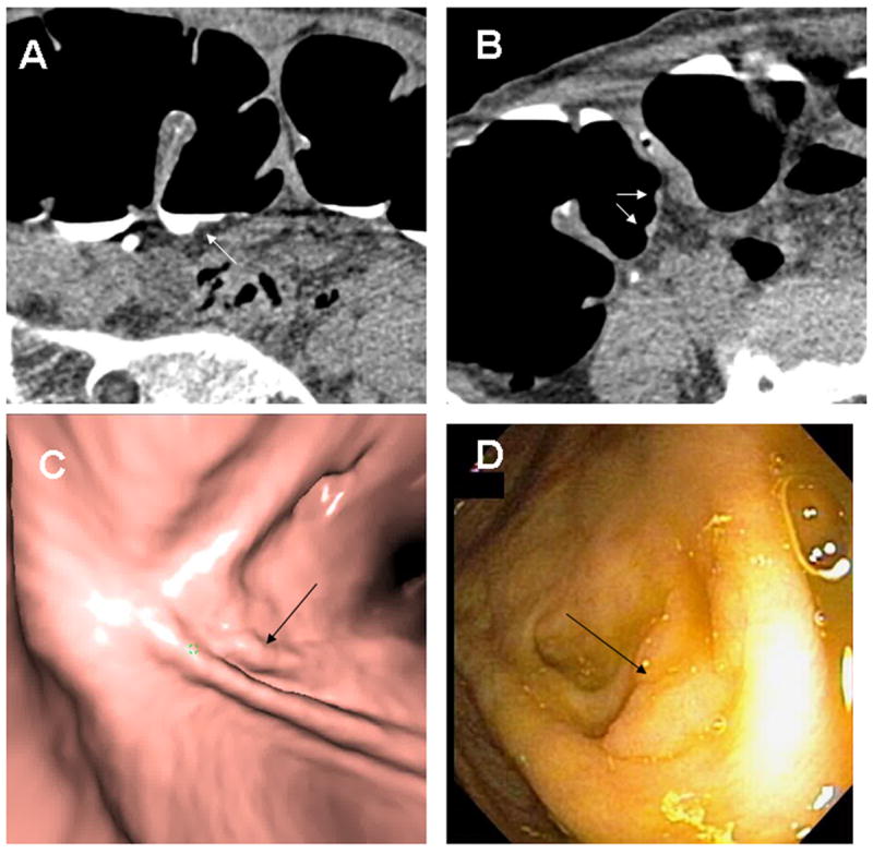
Fig. 2. Cecal Tubular Adenoma.

A 10 × 2 mm tubular adenoma with low-grade dysplasia is seen in retrospect at the base of a fold in the cecum (arrows) on supine 2D (A), prone 2D (B), 3D endoluminal (C) and colonoscopy (D). Note the polyp is submerged in tagged fluid on the supine images and would be obscured on supine endoluminal views without fluid subtraction. The linear configuration could be misinterpreted as a fold however this is immediately adjacent to a normal-appearing fold and only extends a short distance.
