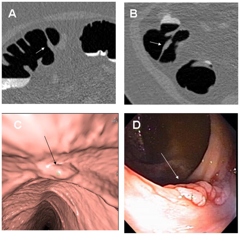
Fig. 4. Ascending Colon Tubular Adenoma.

8 × 3mm tubular adenoma in the ascending colon along a fold (arrows) seen in retrospect on 2D supine (A), 2D prone (B), 3D endoluminal (C) and colonoscopy (D). On the 3D endoluminal views, the less than optimal distension causes some irregularity of the mucosa that potentially could have obscured the detection of the polyp. On the 2D views, the focal enlargement of the fold should raise suspicion for an associated polyp.
