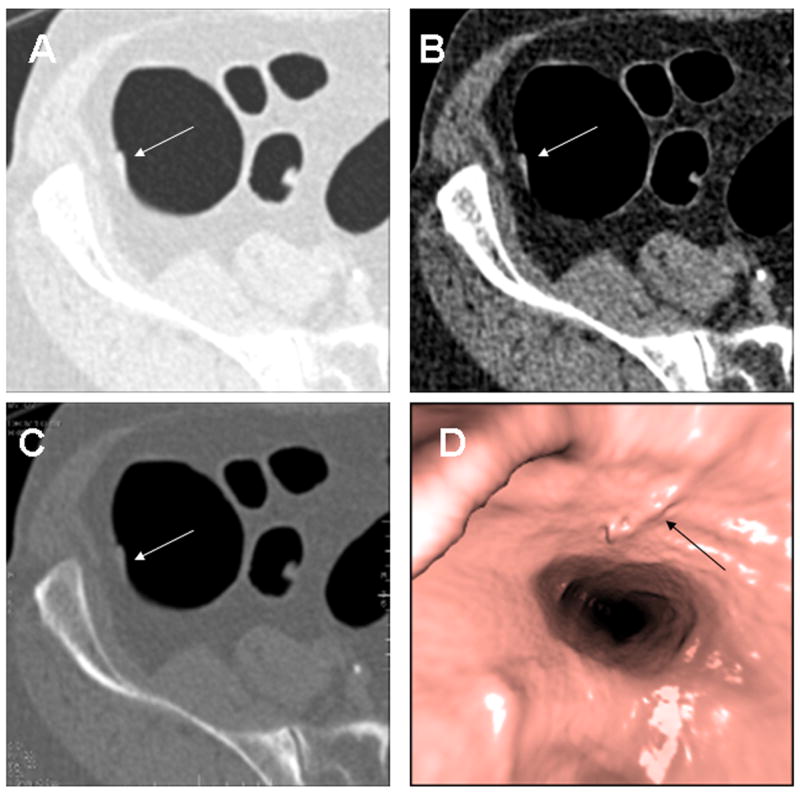
Fig. 5. Cecal Tubulovillous Adenoma.

8 × 2mm low-grade tubulovillous adenoma in the cecum (arrows) seen in retrospect on 2D lung window (A), soft tissue window (B), colon window (C) and 3D endoluminal (D) images. In this example the polyp is seen well on all three window settings.
