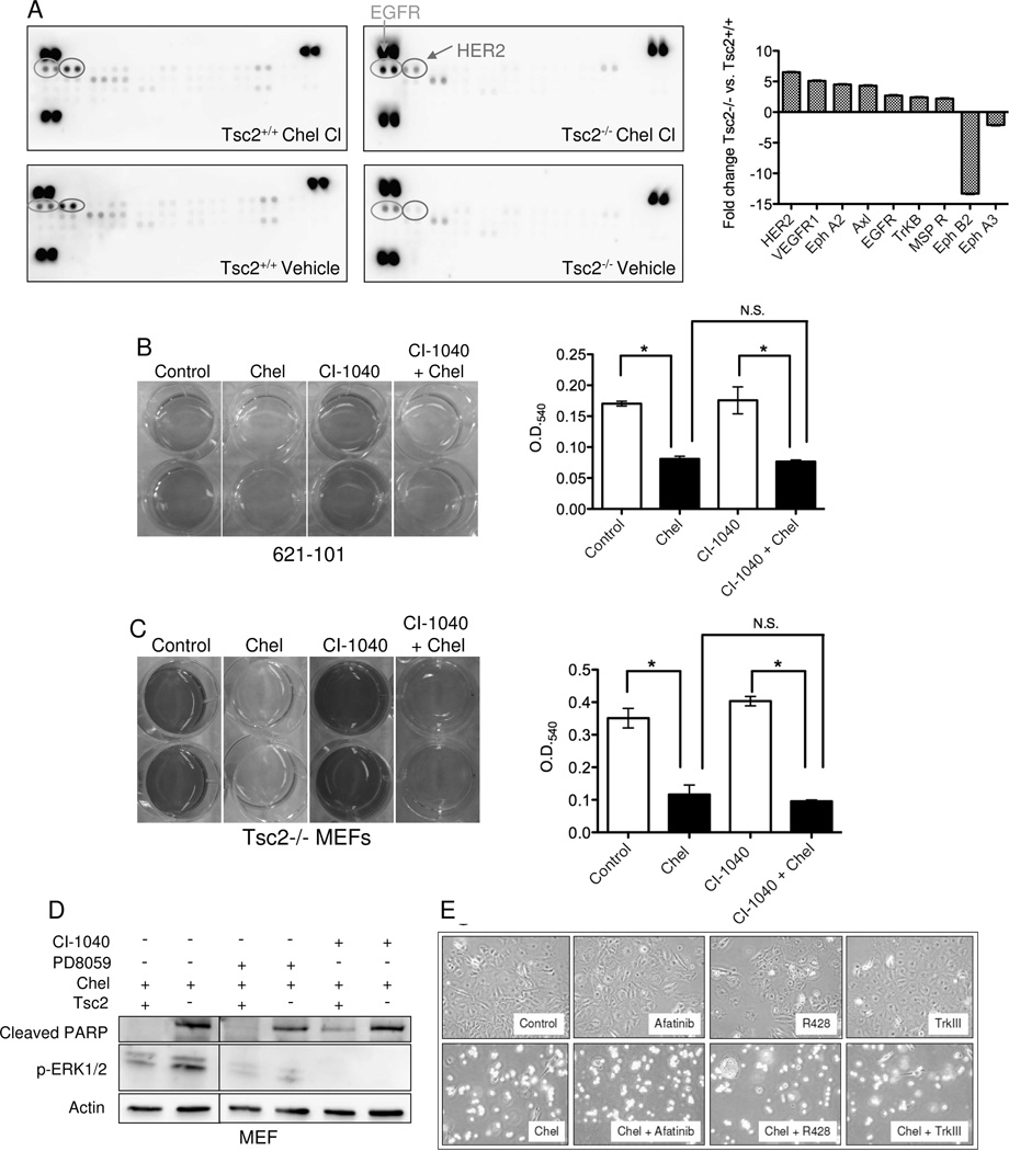
Figure 4. Chelerythrine chloride induces EGFR and HER2 signaling in Tsc2-null cells.
A) Phospho-receptor tyrosine kinase array of Tsc2−/− and Tsc2+/+ MEFs treated with 1 uM chelerythrine chloride for 1h. The bar graph shows densitometric analysis of relative changes in chelerythrine-treated Tsc2−/− vs. Tsc2+/+ cells after normalization to vehicle control. B, C) 621-101 cells and Tsc2−/− MEFs were pretreated with the MEK inhibitor CI-1040 (1 uM) for 16 hours followed by the addition of 5 uM chelerythrine chloride (621-101) or 2 uM chelerythrine (MEFs) for 2 hours. Cell proliferation was measured by crystal violet staining. MEK inhibition did not block the chelerythrine-induced effects in either cell type. (D) Immunoblot analysis of Tsc2+/+ and Tsc2−/− MEFs pretreated with the ERK inhibitor PD98059 (50 uM) or the MEK inhibitor CI-1040 (1 uM) for 16 hours followed by chelerythrine chloride (1 uM) for 3 hours. E) Phase contrast images (4X) of Tsc2-null MEFs treated with chelerythrine chloride (2 uM) and/or the EGFR inhibitor Afatinib (300 nM), the Axl inhibitor R428 (300 nM), or the Trk-beta inhibitor TrkIII (300 nM). Inhibitors were chosen based on results from panel A. Images were captured 24 hours post addition of chelerythine.

