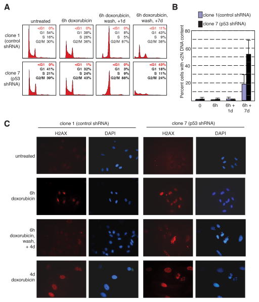
Figure 5.
p53-expressing cells recover from short-term chemotherapeutic treatment whereas p53-ablated cells do not. A–B: The U2OS derivatives clone 1 (expressing control shRNA) or clone 7 (expressing p53 shRNA) were untreated or treated with 0.05 μg/ml doxorubicin for 6 hours, then washed, and allowed to grow in complete medium without drug for 1 or 7 days. DNA content was assayed by propidium iodide staining. Representative histograms from a single experiment are shown (A). Percent of cells with less than 2N DNA content was quantitated. The average of three independent experiments was averaged and is shown as a bar graph (B). C: Both p53-expressing and p53-null cells show activation of the DNA damage checkpoint in response to short-term treatment with a chemotherapeutic agent. Clone 1 (expressing control shRNA) or clone 7 (expressing p53 shRNA) cells were either untreated, treated with 0.05 μg/ml doxorubicin for 6 hours or 4 days, or treated with doxorubicin for 6 hours, washed, and then incubated in the absence of drug for 4 days. Cells were then subjected to immunofluorescence staining for γ-H2AX (left panels, red). Nuclei were counterstained with DAPI (right panels, blue). D: p53-expressing cells recover from a 6 hour treatment with a chemotherapeutic agent, but not after a 2 day treatment. Clone 1 (expressing control shRNA) or clone 7 (expressing p53 shRNA) cells were treated with 0.05 μg/ml doxorubicin for 6 hours or 2 days, then washed, and allowed to grow without drug for 2–7 days. Cells were then double-stained for phosphorylated histone H3 and DNA content (propidium iodide), and analyzed by flow cytometry. Results are plotted as the ratio of treated cells positive for phosphorylated histone H3 compared to untreated cells positive for histone H3 phosphorylation.

