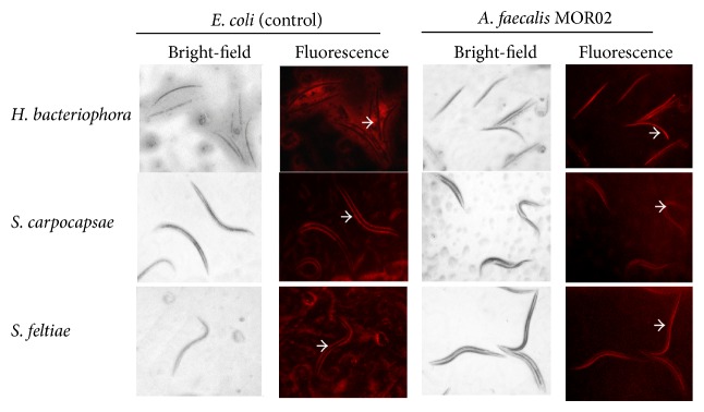
Figure 3.

Association of A. faecalis MOR02 IJ2 nematodes. Bright-field and fluorescence microscopy analyses of the association of E. coli and A. faecalis MOR02-Cherry with H. bacteriophora, S. carpocapsae, and S. feltiae. In E. coli (negative control), fluorescence is observed outside nematodes (white arrows), whereas A. faecalis MOR02 fluorescence is located inside the nematodes (white arrows). Gut autofluorescence of H. bacteriophora is not clearly observed due to red fluorescence of A. faecalis MOR02.
