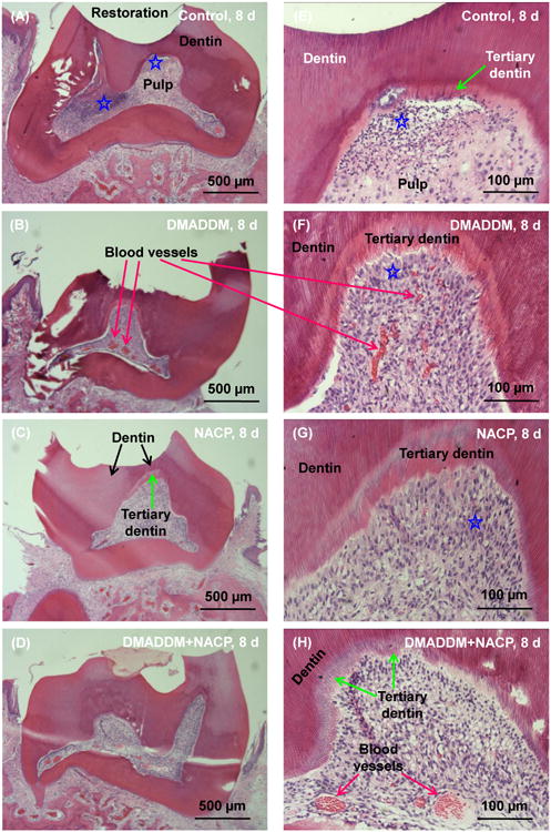
Figure 2.

Histological analyses of rat tooth cavities at 8 d. (A-D) Control group, DMADDM group, NACP group, and DMADDM + NACP group. (E-H) The same four groups at a higher magnification. Control and DMADDM group exhibited disruption of the odontoblast layer associated with a medium inflammatory response in the pulp. Blood vessels were observed, and a thin layer of tertiary dentin could be found. NACP group and DMADDM+NACP group had generally normal pulp tissues, with much thicker tertiary dentin than Control. Stars indicate areas with inflammatory cell infiltration.
