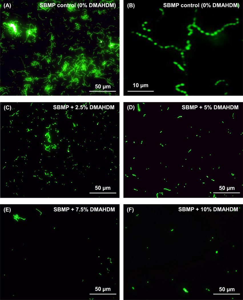
Fig. 3.
Typical fluorescent images of S. mutans colonization on bonding agent resin disks containing DMAHDM: (A) SBMP control, (C) SBMP + 2.5% DMAHDM, (D) SBMP + 5% DMAHDM, (E) SBMP + 7.5% DMAHDM, (F) SBMP + 10% DMAHDM. A higher magnification for SBMP control is shown in (B). S. mutans were incubated on resin disks for 4 h. S. mutans had long chains in (A) which is more clearly shown in (B) on SBMP control, versus individual bacteria dots at 10% DMAHDM in (F).

