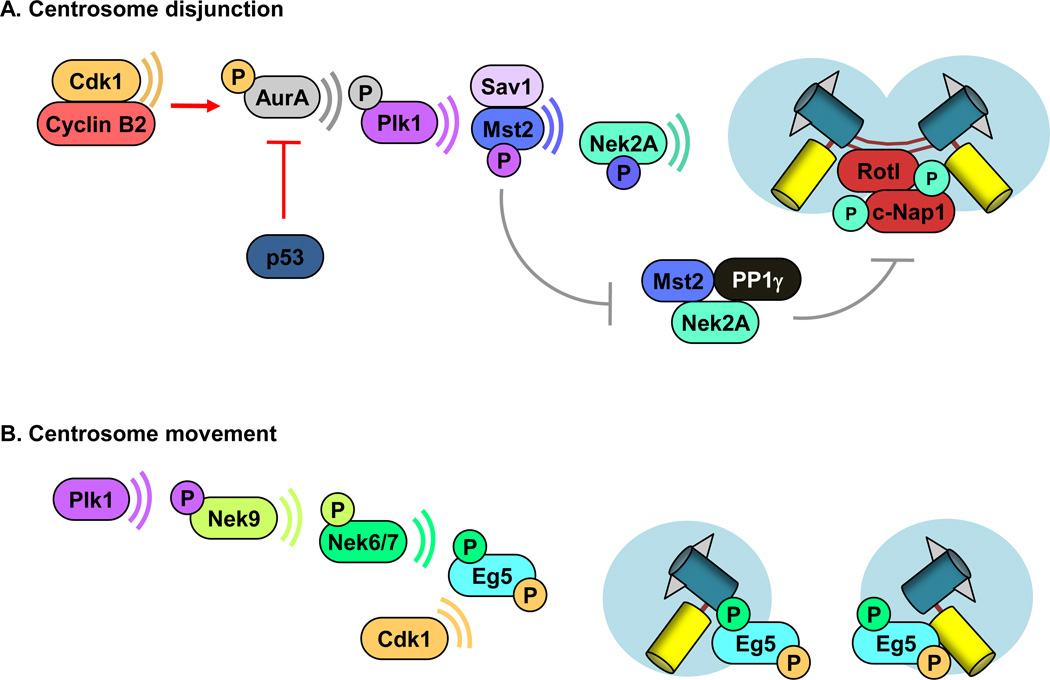
Figure 1. Centrosome dynamics.
Centrosome separation occurs in two parts from late G2 through mitosis: disengagement and movement. (A) Hypothetical model for centrosome disjunction. Centrosome-associated cyclin B2/Cdk1 acts to initiate centrosome disjunction through phosphorylation of Aurora A, which in turn phosphorylates Plk1. Activated Plk1, phosphorylates Sav1-bound Mst2, triggering Mst2-mediated phosphorylation of Nek2A. Activated Nek2A phosphorylates the centrosome linker proteins C-Nap1 and rootletin, causing the physical separation of sister centrosomes. PPlγ antagonizes Nek2A phosphorylation of C-Nap1 and Rootletin. [13, 52, 57]. (B) Model for centrosome movement. Plk1 targets Eg5 to centrosomes through sequential phosphorylation of Nek9 and Nek6/7. Cdk1 has also been proposed to be important for Eg5 activation and binding to microtubules [13, 57, 63, 93].

