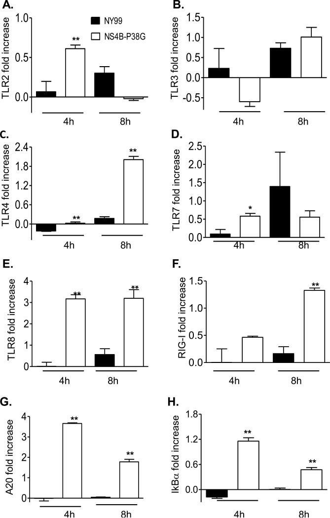
Fig. 5.
PRR and NF-κB -related gene expression in WNV-infected THP-1 cells. THP-1 cells were infected with WNV NY99 and WNV NS4B-P38G mutant at a MOI of 0.5. TLRs 2, 3, 4, 7 and 8 (A- E), RIG-I (F), A20 (G), and IκBα (H) gene expression at 4 and 8 hrs post infection were determined by using a Q-PCR assay. The fold increase compared to that in the mock-infected group is shown. Data are presented as means ± SEM, n = 4. * P < 0.05 or **P < 0.01 for NY99 vs. NS4B-P38G. Results presented are one representative of two similar experiments.

