
Figure 5.
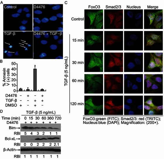
CKIinhibition blocks TGF-β-induced apoptosis in Hep3B cells. (A) CKI-ε inhibition blocks TGF-β-induced apoptosis. Cells were treated with CKI inhibitor D4476 (10 μmol/L), TGF-β (5 ng/mL) or D4476 plus TGF-β for 48 h. Apoptosis was assessed by staining with Hoechst 33342 for nucleus condensation as described in ‘MATERIALS AND METHODS’. A representative field of cells with the indicated treatments has been shown. Typically apoptotic cells with apoptotic nuclei were marked with white arrows. (B) Cells were treated with various conditions as indicated. Apoptosis was assessed by FACS analysis based on Annexin V-PI double staining. Statistical analysis was performed to assess the ratio of apoptosis. Data represent the mean values of three independent experiments (*P < 0.05). (C) D4476 abolishes TGF-β-stimulated Smad2/3 and FoxO3 translocation. Hep3B cells grown on cover slips were pretreated with D4476 for 30 min and continuously incubated with TGF-β (5 ng/mL) or not. Immunofluorescence double staining was performed to evaluate the presence of Smad2/3 and FoxO3 with antibodies specifically recognizing Smad2/3 and FoxO3. The bound Smad2/3 primary antibody was visualized with Cy3-conjugated donkey anti-mouse IgG (Red) and FoxO3 was with FITC-conjugated goat anti-rabbit IgG (Green). Nuclei were stained with specific dye DAPI (blue). (D) D4476 blocks TGF-β-induced Bim up-regulation. Cells were pretreated with D4476 for 30 min followed by TGF-β (5 ng/mL) incubation for various times ranging from 0 to 720 min. Cells were harvested and cell lysates were prepared for Western blotting to detect levels of Bim and Bcl-xL with specific antibodies recognizing Bim and Bcl-xL. Equal protein loading was determined by β-Actin. Relative band intensities (RBIs) were analyzed by the Image J software. Representative bands were shown. Each experiment was conducted in triplicate and repeated twice independently