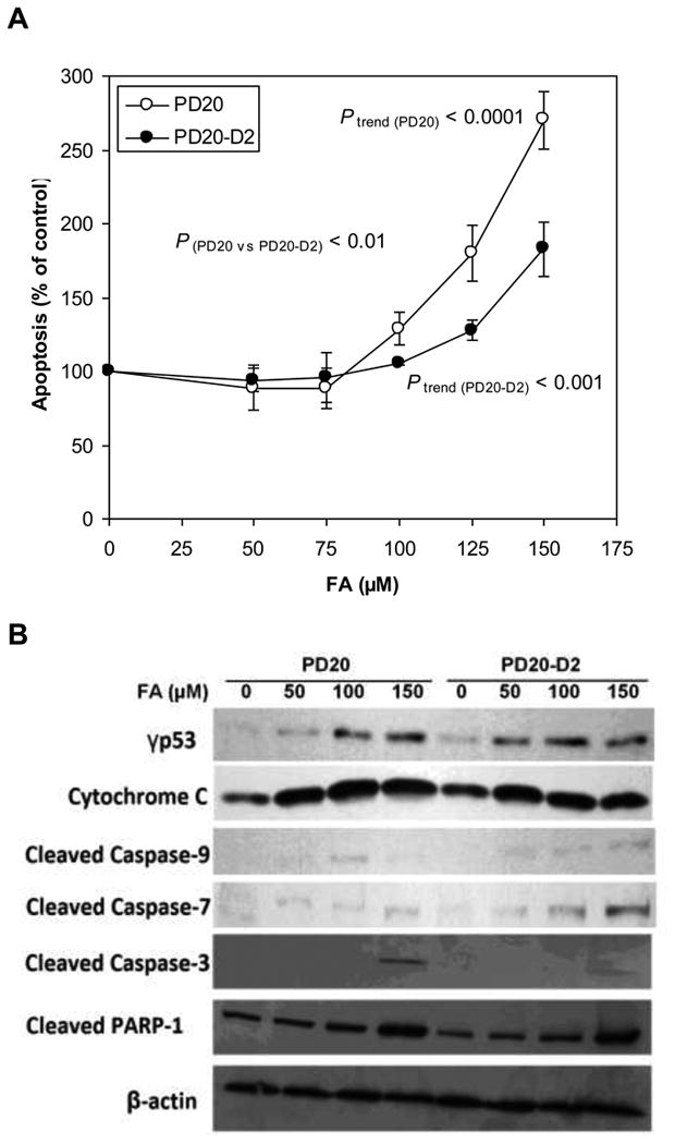
Fig. 5.

Apoptosis induced by FA in PD20 and PD20-D2 cells.
(A) Apoptosis in FA-treated PD20 and PD20-D2 cells as a percentage of untreated control cells. FA induced apoptosis in a dose-dependent manner in both cell lines (Ptrend (PD20) < 0.0001 and P trend (PD20-D2) < 0.001) with greater effects in PD20 cells (P(PD20 vs PD20-D2) < 0.01).
(B) Apoptosis-related protein expression in FA-treated PD20 and PD20-D2 cells as a percentage of untreated control cells. p53 was activated and caspase signaling pathway was induced by FA at similar levels in both PD20 and PD20-D2 cells. β-actin is used as loading control.
