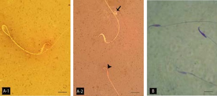
Fig 5.
Light microscopic architecture of sperms; (A-1) normal sperm with unstained cytoplasm and (A-2) morphologically abnormal sperm with cytoplasmic droplet on the tail piece (arrow) and the dead sperm with stained cytoplasm (Head arrow). (B) Two nuclear immature sperms with faint stained nucleuses and one nuclear matured sperm on top right hand side are showed. A-1 and A-2, Eosin-negrosin and B, Aniline-blue staining, 1000×.

