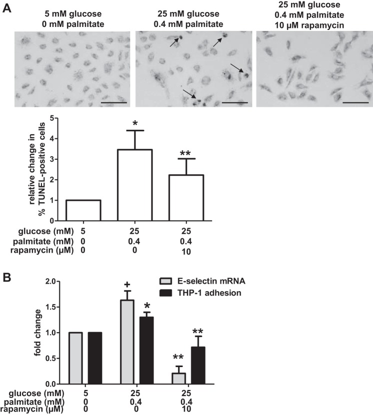
Fig. 3.
Glucose and palmitate increase apoptosis and inflammation. A: HAECs were incubated in media containing the indicated concentrations of glucose, palmitate, and rapamycin. TUNEL staining was used to label DNA strand breaks, and they were visualized under brightfield microscopy. Punctate nuclear staining (arrows) indicates DNA strand breaks. Bar, 100 μm. The percentage of cells containing punctate nuclear staining (“TUNEL-positive”) was calculated for each treatment group (n = 5 for control; n = 5 for excess nutrient; n = 3 for excess nutrient+rapamycin). The graph indicates that excess nutrients increased the percentage of TUNEL-positive cells and that treatment with rapamycin attenuated these effects of glucose and palmitate on TUNEL staining. B: incubation of HAECs in media containing the indicated concentrations of glucose and palmitate also increased mRNA levels of the proinflammatory cytokine E-selectin (n = 3) and adhesion of THP-1 monocytes (n = 9) relative to control-treated cells. Treatment with rapamycin attenuated these changes. For A and B, +P = 0.07 and *P ≤ 0.05 compared with control conditions; **P < 0.05 compared with excess nutrient conditions.

