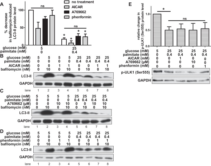
Fig. 5.
Chemical activation of AMPK does not restore autophagy in HAECs exposed to glucose and palmitate. A: bafilomycin-induced LC3-II protein levels (relative to loading control) were measured by Western blot in HAECs incubated with 1 mM AICAR, 10 μM A769662, 1 mM phenformin, or no activator. Relative to cells incubated in control conditions, the percent decrease in LC3-II protein level is shown for cells incubated in nutrient excess conditions (n ≥ 3). *P < 0.05 relative to control conditions; ns, no significant effect of AMPK activators within each treatment condition. B–D: LC3-II and GAPDH protein bands are shown on representative Western blots for AICAR (B), A769662 (C), and phenformin (D) treatments. E: the relative change (after adjusting for loading control) in protein level of phosphorylated ULK1 (p-ULK1 Ser555), an autophagy-activating site targeted by AMPK, is shown (n ≥ 4). A representative Western blot is shown for p-ULK1 (Ser555) and GAPDH below. *P < 0.05; ns, no significant effect of AMPK activators in the excess nutrient condition.

