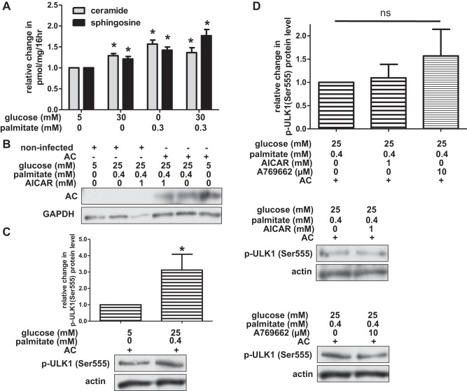
Fig. 6.
Overexpression of acid ceramidase (AC) restores ULK1 phosphorylation. A: human umbilical vein endothelial cells (HUVECs) were incubated in media containing glucose and/or palmitate for 16 h, and ceramide and sphingosine levels were measured using [3H]-l-serine (adjusted for protein content). Data are expressed as change in ceramide or sphingosine levels for each treatment group relative to control-treated cells (n ≥ 3). B: noninfected HAECs and HAECs infected with acid ceramidase lentivirus were incubated in media containing glucose, palmitate, and AICAR. Acid ceramidase expression was evaluated by Western blotting. C: HAECs ectopically expressing acid ceramidase were incubated in control or nutrient excess conditions. Densitometric analysis of p-ULK1 (Ser555) protein level (relative to loading control) is shown (n = 6) with representative Western blot below. D: HAECs ectopically expressing acid ceramidase were incubated in media containing high concentrations of glucose and palmitate as well as AICAR or A769662. Densitometric analysis of p-ULK1 (Ser555) protein level (relative to loading control) is shown (n = 3) with representative Western blots below. For A, C, and D, *P < 0.05 compared with control conditions; ns, no significant difference.

