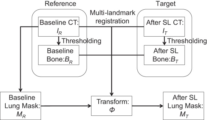
Fig. 1.

The pipeline of automated segmentation. The process of lung segmentation is shown, starting with thresholding the computed tomography (CT) images to obtain the bone mask. A multilandmark registration was then computed using both the pair of CT images and the pair of bone masks (after distance transform for the deformable registration). The resulting lung mask after saline lavage (SL) was computed by applying the transform to the baseline lung mask. IR, reference image; IT, target image; BR, bone mask for the reference IR; BT, bone mask for the target SL image IT; MR, binary lung mask obtained from the IR; MT, binary lung mask obtained from the IT.
