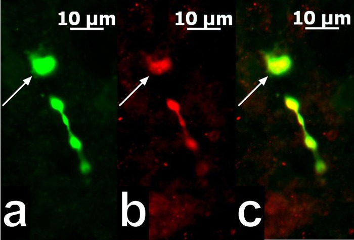
Fig. 3.
A color photomicrograph of an ARC proopiomelanocortin (POMC) neuron from which an electrophysiological recording was taken. a: Biocytin labeling of the neuron filled throughout the recording and visualized with AF488. b: Immunoreactivity for β-endorphin located in the soma and varicosities of the cell in a as visualized with AF546. c: Merged photomicrographs from a and b illustrating the ARC neuron (arrows) that was double-labeled with biocytin and β-endorphin immunoreactivity.

