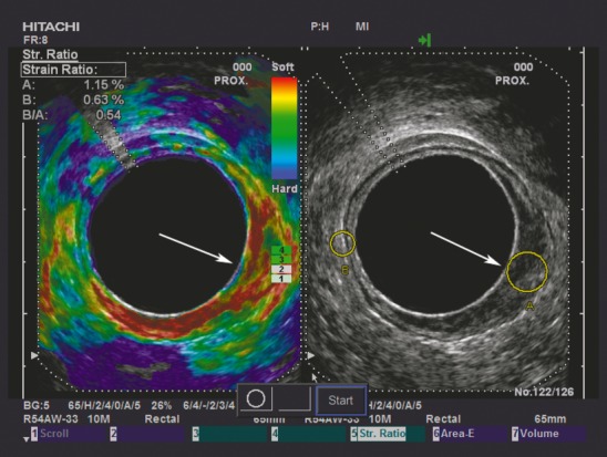
Figure 1.

Split-screen image shows a B-mode image with strain ratio (SR) regions of interest on the right and an elastogram on the left. The tumour is situated from 2 to 7 o'clock (white arrow). The tumour appears softer (more red) than the same-depth reference tissue on the elastogram, and the SR (B/A) with SR measurement indicative of an adenoma (SR = 0.54) is displayed in the upper left-hand corner. When evaluated ultrasonographically it is difficult to determine whether the tumour is an early adenocarcinoma or an adenoma, as the hypoechoic mucosal layer is not clearly distinguished from the hyperechoic submocosal layer in the tumour region. The resection specimen was histopathologically confirmed as adenoma.
