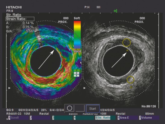
Figure 2.

To contrast the adenoma in Fig.1, Fig. 2 demonstrates an adenocarcinoma situated from 11 to 3 o'clock, as indicated by the white arrow. The tumour appears harder (more blue) than the same-depth reference tissue on the elastogram, and a strain indicative of an adenocarcinoma (SR = 5.56) is displayed in the upper left-hand corner.
