
Fig 2.
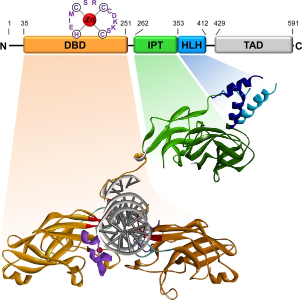
Structure of EBF1. A schematic presentation of the domain structure of murine EBF1 [modified after Hagman and Lukin 38] and the crystal structure of a DNA-bound EBF1 dimer that lacks the C-terminal transactivation domain [modified after Treiber et al. 31 using PDB file 3MLP are depicted]. The structure was modeled using Discovery Studio 3.5 Visualizer, Accelrys Inc., San Diego, CA. The DNA-binding domain (DBD) is colored in orange, the IPT domain in green, the helix-loop-helix (HLH) dimerization domain in blue and the C-terminal transactivation domain (TAD) in light gray. The DNA interacting modules within the DBD are colored in red (central motif), purple (zinc coordinating motif) and turquoise (GH loop). Zinc ions are represented by red spheres. The border amino acids of each domain are indicated in the schematic.